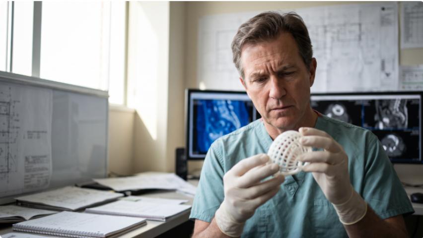
The Limits of Conventional Implants in Severe Acetabular Defects
Standard implants were never designed for the worst-case scenario — and in revision hip surgery, the worst case is increasingly common.
When bone loss is severe enough, off-the-shelf solutions stop being solutions at all. Paprosky Type III defects, classified as the most destructive pattern in revision total hip arthroplasty, present surgeons with near-total loss of the acetabular rim, compromised column integrity, and geometry that no catalog implant can reliably match. The anatomy is unpredictable by definition — no two defects erode identically, and the remaining bone that must anchor a new implant varies dramatically from patient to patient.
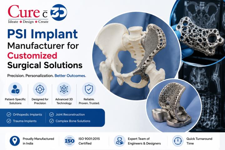
Standard triflange constructs and jumbo hemispherical cups attempt to compensate through size and screw fixation, but they carry a fundamental limitation: they're built to fit a population average, not an individual pelvis. In practice, this means surgeons must accept imperfect contact, reduced load distribution, and elevated rates of aseptic loosening. What typically happens is that micro-motion at the implant-bone interface prevents the biological ingrowth that long-term fixation depends on.
This clinical reality has driven a measurable shift toward patient-specific implants designed through digital planning — a precision-first mindset that starts with the patient's CT data rather than a standard size chart. Emerging data on custom 3D-printed acetabular implant clinical outcomes suggest this approach is reshaping what's achievable in the most complex reconstructions, with research showing implant survival rates that standard constructs rarely match in equivalent defect severity.
What those outcomes actually look like over a nine-year horizon — and why porous lattice design is central to the story — is where the evidence gets compelling.
Long-Term Clinical Outcomes for Custom 3D-Printed Acetabular Implants
Custom 3D-printed titanium implants are delivering survival rates that challenge what orthopedic surgeons once considered achievable in the most compromised acetabular anatomy.
The headline number is hard to ignore: a 9-year survival rate of 96.3% for custom titanium constructs in severe acetabular defects, according to data published by the Journal of Bone and Joint Surgery. For a patient population defined by catastrophic bone loss — where conventional augments routinely fail within a decade — that figure represents a fundamental shift in expectations.
Porous lattice structures are central to explaining why these implants perform so well over time. Unlike smooth-surfaced components that rely on cement or screw fixation alone, lattice geometries mimic the trabecular architecture of cancellous bone, creating a scaffold that actively invites osseointegration. Research published in PMC on porous metal implants confirms that this biological ingrowth — rather than mechanical fixation alone — is what drives long-term stability. In practice, the most meaningful stabilization window closes within the first postoperative year, after which bone-implant integration tends to plateau at durable, load-bearing levels.
Functional recovery in patients with severe bone loss has also proven more substantial than historical benchmarks suggested. Hip scores, pain indices, and mobility metrics all show meaningful improvement at intermediate follow-up, a pattern consistent with findings from acetabular reconstruction outcome studies. Understanding how digital pre-operative planning drives these results helps clarify why outcomes differ so sharply from standard-implant cohorts.
The same precision engineering that produces these acetabular results is now being applied to an entirely different anatomical challenge — and the stakes in spinal trauma surgery are, if anything, even higher.
Navigating Complex Spinal Trauma with 3D-Printed Surgical Guides
Patient-specific surgical guides are transforming spinal reconstruction with the same precision logic driving advances in acetabular reconstruction — and nowhere is that more apparent than in cervical spine trauma.
Freehand screw placement in the cervical spine is one of surgery's most unforgiving technical demands. The anatomy is compact, the margins for error are measured in millimeters, and a misplaced pedicle screw can mean neurological injury. In fracture cases — where normal anatomical landmarks are distorted — the difficulty compounds significantly. Surgeons have historically relied on tactile feel and intraoperative fluoroscopy to navigate, an approach that works in experienced hands but remains inherently variable.
3D-printed surgical guides eliminate that variability by translating virtual plans into physical reality on the table. Based on preoperative CT imaging, patient-specific guide templates lock onto bone surfaces and direct screw trajectories with pre-engineered precision — the same principle behind preoperative 3D surgical planning that is now standard in complex reconstruction. According to research published in PMC, this approach yields measurable results across spinal applications:
- Patient-specific 3D-printed drill guides achieve 99.5% accuracy in pedicle screw placement for adult spinal deformity reconstruction, per the International Journal of Spine Surgery
- Accuracy holds even in Type C and Type D pedicles — the most challenging morphologies where freehand techniques carry the highest breach risk
- Guide-assisted placement significantly reduces cortical perforation rates compared to freehand or fluoroscopy-only methods
- Preoperative planning enables screw length and diameter optimization before the patient enters the OR
The same workflow that addresses Paprosky type III acetabular defects' 3D printing challenges — digitizing complexity, engineering a patient-specific solution, then executing with precision — applies directly here. What once demanded rare surgical artistry is becoming a reproducible, data-driven process that any well-trained surgical team can execute consistently. That shift from art to engineering carries implications that extend well beyond accuracy alone — including measurable effects on operative time and patient exposure to radiation, which the next section examines in detail.
Quantifying the Efficiency: OR Time and Fluoroscopy Reduction
3D-printed surgical guides deliver measurable, case-level efficiency gains that matter equally to patient safety teams and hospital finance departments.
The numbers are hard to ignore. According to a PRISMA scoping review, 3D-printed guides for cervical spine fractures reduced operative time by an average of 32.3 minutes per case — a figure that compounds dramatically across a busy surgical program. The same review found that 3DP-assisted surgery cut intraoperative blood loss by a mean difference of 121.6 ml and reduced fluoroscopy frequency by 3.8 shots per case. Each of those metrics represents a direct, quantifiable improvement in patient outcomes.
Reduced blood loss directly improves safety margins. Saving over 100 mL intraoperatively lowers transfusion risk, supports faster recovery, and reduces ICU burden — benefits that extend well beyond the OR. For patients already managing compromised anatomy, including those receiving patient-specific implants for severe acetabular bone loss, minimizing surgical stress during reconstruction is critical. Fewer fluoroscopy shots also translate to meaningfully lower radiation exposure for both patient and surgical staff over time, a factor that hospital safety officers increasingly track.
The economic case is equally compelling. Thirty-plus minutes of reclaimed OR time — at an average cost of $36–$37 per minute in US hospitals — represents real budget impact per procedure. Understanding how pre-operative planning reduces intraoperative risk is increasingly central to that equation. These efficiency gains are precisely what drive surgeons to advocate for 3D-printed guides — a perspective worth examining closely.
The Surgeon's Perspective: From Artistry to Millimeter Precision
Patient-specific technology doesn't just change what surgeons can do — it changes how confidently they can do it. The efficiency gains documented in previous sections are real, but they tell only part of the story. Equally significant is the psychological shift surgeons describe when entering an operating room with a plan built around one patient's exact anatomy rather than a population average.
Dr. Ryan Goodwin of Cleveland Clinic captures this directly: "FIREFLY® patient-specific, 3D printed drill guides have dramatically reduced surgical times while improving accuracy in pedicle screw placement in very complex anatomic situations." The phrase "complex anatomic situations" is key. These are cases — severe deformities, failed prior surgeries, significant bone loss — where standard instrumentation simply doesn't fit. For procedures like revision total hip arthroplasty 3D printed custom triflange reconstruction, where bone stock is unpredictable, and margins for error are vanishingly thin, that confidence isn't a soft benefit. It's a clinical necessity.
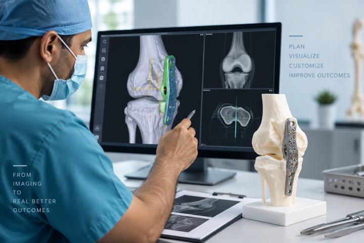
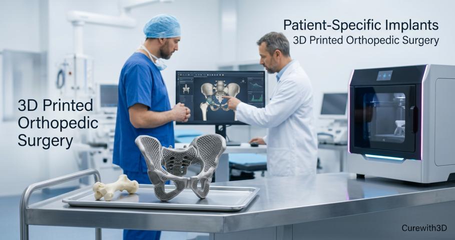
Virtual surgical planning (VSP) is what makes that confidence possible before the patient reaches the OR. By simulating the procedure digitally — positioning implants, anticipating anatomical obstacles, rehearsing the surgical sequence — teams arrive with a shared blueprint that bridges the gap between engineering precision and clinical judgment. Understanding how pre-operative planning works in practice helps illustrate why surgeons consistently describe VSP as transformative rather than supplementary.
On the other hand, this workflow requires genuine collaboration. Engineers need clinical context; surgeons need to engage with design parameters. When that partnership functions well, the result is a custom guide or implant that feels less like an external tool and more like an extension of the surgeon's own preparation — converting what was once artistry into repeatable, measurable precision. The outcomes behind that shift deserve closer examination in the takeaways ahead.
What You Need to Know: Key Takeaways for Surgical Teams
Patient-specific 3D implants and printed surgical guides have moved from experimental to evidence-backed — and the data across multiple domains tell a consistent story of improved precision, safety, and efficiency.
The clinical evidence now makes a compelling case for patient-specific approaches as a new standard in complex reconstruction. Here's what surgical teams should carry forward from the research:
- 96%+ implant survival in severe bone loss cases. Custom acetabular implants designed for Paprosky III defects demonstrate survival rates exceeding 96% at mid-term follow-up, according to JBJS research on custom acetabular reconstruction — outcomes that off-the-shelf hardware rarely matches in comparable defect severity.
- 32 minutes saved per spinal trauma case. As covered earlier, 3D-printed guide templates consistently reduce OR time in spinal procedures, with time savings that compound across high-volume centers.
- 99.5% pedicle screw accuracy. Evidence from cervical spine trauma studies confirms that custom guides reduce freehand placement error to near-negligible levels.
- Long-term structural stability. Per JBJS, 3D-printed constructs stabilize through osseointegration with negligible migration after the first year — a critical marker for durable reconstruction outcomes.
- Reduced radiation and intraoperative variability. Fewer fluoroscopy passes and less reliance on real-time correction translate directly into lower exposure risk for both patient and OR team.
For teams managing complex reconstructions, predictable surgical approaches built on patient-specific planning aren't a luxury upgrade — they're a risk-reduction strategy with measurable returns at every stage of care.
Implementing Patient-Specific Solutions with Curewith3D
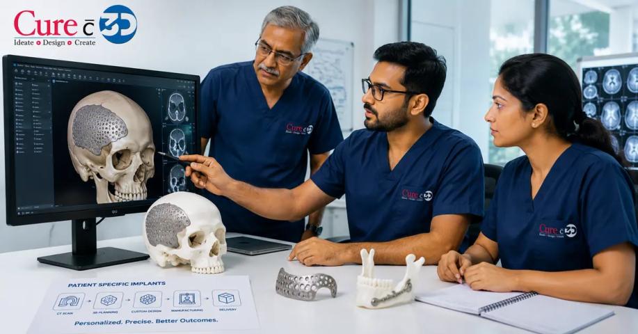
Patient-specific 3D implants are only as effective as the engineering and clinical expertise behind them — and that gap between design intent and surgical outcome is exactly where Curewith3D operates. The platform bridges computational precision with real-world anatomy, translating patient imaging data into millimeter-perfect surgical guides and biocompatible implants built for each individual case.
Biocompatible materials aren't a checkbox — they're the foundation of long-term outcomes. Every implant must integrate with living tissue, tolerate mechanical load, and resist degradation over decades. Research published in Applied Sciences confirms that porous titanium constructs engineered to patient-specific geometry consistently support osseointegration while preserving structural integrity — properties that standard off-the-shelf components simply cannot replicate across complex anatomical presentations.
The broader shift happening across orthopedic reconstruction is a move toward surgical predictability as a standard of care, not a premium exception. When virtual planning precedes the OR and implants arrive pre-matched to the defect, surgeons spend less time problem-solving and more time executing. The distal femoral reconstruction case in Curewith3D's portfolio illustrates this directly — a porous titanium implant matched precisely to native bone geometry, designed before the first incision.
For surgical teams managing complex reconstruction cases — tumor resection, revision arthroplasty, or high-demand trauma — the path forward starts with a consultation. Contact Curewith3D to explore how virtual surgical planning and patient-specific manufacturing can bring that level of precision to your next case.