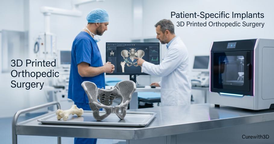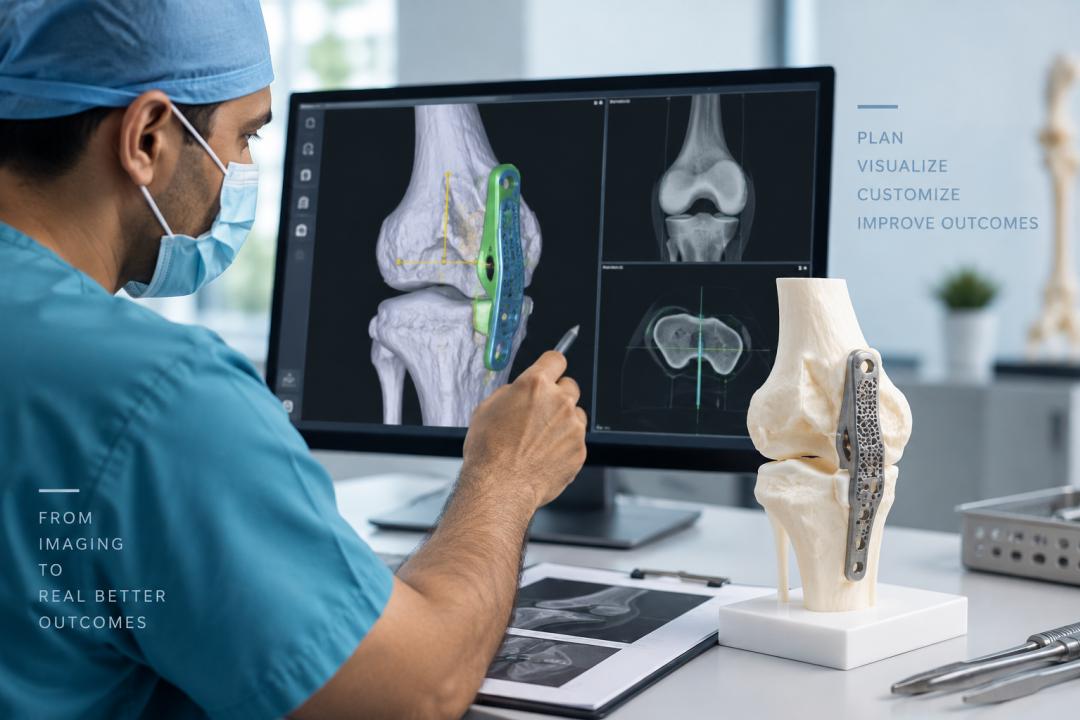
Modern healthcare is increasingly moving toward digital technologies that help medical professionals understand complex anatomy, prepare for procedures, and make more informed decisions before entering the operating room. Among these technologies, virtual surgery planning and 3D anatomical models are becoming valuable tools for surgical preparation, medical education, and patient-specific treatment planning.
These technologies combine medical imaging, digital modeling, and advanced visualization to create a more detailed understanding of individual anatomical structures. Rather than relying only on conventional two-dimensional scans, healthcare professionals can use digital and physical models to study anatomy from different perspectives and prepare for challenging procedures.
What Is Virtual Surgery Planning?
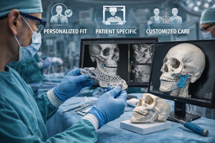
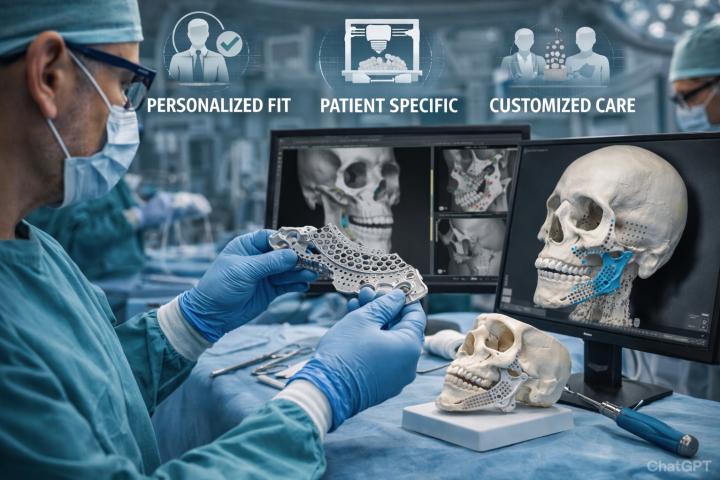
Virtual surgery planning is a digital approach that allows surgeons and medical teams to plan a procedure before it takes place. It generally begins with patient-specific medical imaging data, such as CT or MRI scans, which can be processed to create a three-dimensional representation of the relevant anatomy.
This digital model enables the surgical team to examine anatomical structures, evaluate the surgical site, and explore potential approaches before the actual procedure. Depending on the clinical application, virtual planning may include anatomical segmentation, 3D reconstruction, measurement, digital simulation, and planning of implant or guide placement.
For a more detailed explanation of the concept and its clinical relevance, virtual surgery planning can provide further insight into how this technology is integrated into modern surgical workflows.
How Virtual Surgery Planning Works
The virtual planning process typically involves several stages. Although the exact workflow can differ depending on the procedure, the general process includes:
1. Medical Imaging
High-quality patient imaging provides the foundation for the digital planning process. CT or other suitable imaging data can be used to visualize bones, anatomical structures, and the target surgical region.
2. Digital Anatomical Reconstruction
The imaging data is processed to create a three-dimensional digital representation. Segmentation techniques may be used to separate relevant anatomical structures from surrounding tissues.
3. Surgical Analysis and Simulation
Surgeons and engineers can then analyze the digital model and evaluate different surgical approaches. Measurements, reference points, implant positions, and other procedural considerations can be assessed during this stage.
4. Development of Patient-Specific Solutions
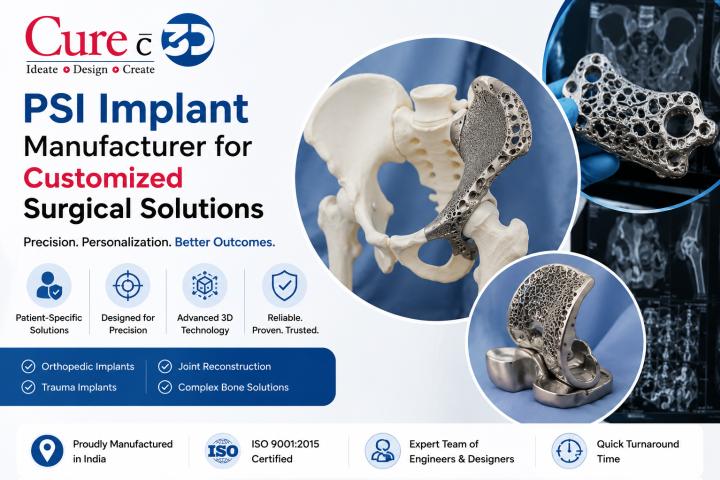
When necessary, the digital plan can support the design of patient-specific implants, cutting guides, drilling guides, positioning guides, and other surgical tools.
5. Clinical Review
The proposed plan can be reviewed and adjusted before treatment. This collaborative process allows surgeons and technical teams to evaluate the plan and make changes where clinically appropriate.
Why Digital Surgical Planning Matters
Complex procedures can involve challenging anatomy, previous surgical changes, bone defects, or structures that are difficult to understand using conventional imaging alone. Digital surgical planning can provide an additional layer of visualization before treatment begins.
Some potential advantages include:
- Better visualization of complex anatomy
- More structured preoperative preparation
- Improved communication between multidisciplinary teams
- Patient-specific planning based on medical imaging
- Opportunity to evaluate surgical strategies before the procedure
- Support for the design of customized surgical tools and implants
Virtual planning does not replace clinical judgment. Instead, it can serve as a digital support system that helps surgeons evaluate anatomy and prepare for procedures in greater detail.
The Role of 3D Anatomical Models
While digital environments provide excellent visualization, physical models can offer another useful perspective. A 3D anatomical model represents patient anatomy in a tangible form, allowing healthcare professionals to inspect structures manually and from multiple angles.
These models can be particularly useful when anatomy is complex or when spatial relationships are difficult to understand through a screen alone.
A physical model may help a surgical team examine areas of interest, identify anatomical landmarks, assess defects, and discuss potential approaches. Models can also be useful outside the operating room, particularly in medical education and professional training.
How 3D Printed Models Support Healthcare Professionals
Advances in medical 3D printing have made it increasingly practical to transform digital anatomical data into physical models. Depending on the intended application, models can be produced using different materials and printing technologies.
Healthcare professionals may use 3D printed anatomical models for several purposes, including:
Surgical Preparation
A physical anatomical model can help surgeons study the geometry and spatial relationships of complex structures before surgery. This can be especially useful in reconstructive or anatomically challenging cases.
Medical Education
Students and trainees can use 3D models to better understand anatomy by examining structures from different orientations. Compared with static textbook illustrations, physical models offer an interactive way to study three-dimensional relationships.
Team Communication
A model can provide a common visual reference for surgeons, engineers, clinicians, and other members of a healthcare team. This may make discussions about anatomy and planned procedures more straightforward.
Patient Communication
In appropriate clinical settings, physical models can also help explain anatomical conditions and treatment concepts to patients. Visual communication may make complex medical information easier to understand.
Combining Digital Planning With Physical Visualization
One of the strongest aspects of modern medical 3D technology is the ability to combine virtual and physical workflows.
A digital model may be used to evaluate anatomy, simulate surgical strategies, and develop patient-specific designs. Once the planning stage is complete, selected digital information can potentially be used to create a physical anatomical model or surgical guide.
This creates a connected workflow:
Medical Imaging → 3D Digital Reconstruction → Virtual Planning → Physical Model or Surgical Tool
The combination of these technologies can provide healthcare teams with both digital and tactile perspectives. Digital planning offers flexibility and precise visualization, while physical models provide a hands-on representation of the patient's anatomy.
Applications in Modern Surgical and Medical Practice
Virtual surgical planning and anatomical modeling can be relevant across various medical specialties. Potential applications include orthopedic procedures, cranio-maxillofacial reconstruction, neurosurgical planning, trauma cases, dental procedures, and reconstructive surgery.
In orthopedics, these technologies can support the evaluation of complex bone defects and the planning of customized solutions. In cranio-maxillofacial surgery, 3D representations can assist with understanding irregular anatomy and reconstruction requirements.
Dental and maxillofacial applications may also benefit from digital planning and 3D visualization when anatomical precision is important. Similarly, medical institutions can incorporate physical models into teaching programs, simulation-based learning, and professional training.
The specific usefulness of these technologies depends on the procedure, imaging quality, clinical objectives, and the expertise of the healthcare team.
The Future of Digital Surgical Planning
As healthcare technology continues to develop, digital surgical planning is expected to become increasingly integrated with advanced imaging, additive manufacturing, computer-aided design, and other digital tools.
Future workflows may involve more sophisticated anatomical segmentation, automated design assistance, improved simulation capabilities, and more seamless connections between planning software and manufacturing systems.
Artificial intelligence and advanced computational methods may also contribute to image processing and planning workflows. However, clinical oversight will remain essential because surgical decisions must be based on patient-specific anatomy, medical evidence, professional expertise, and the overall clinical situation.
Conclusion
Virtual surgery planning and 3D anatomical models represent an important shift toward more personalized and digitally supported healthcare. By combining patient-specific imaging, three-dimensional visualization, and advanced manufacturing technologies, medical teams can gain additional tools for preparation, education, communication, and procedural planning.
As these technologies continue to mature, their role in modern healthcare may expand further. The most meaningful value comes from integrating digital tools into well-structured clinical workflows where technology supports, rather than replaces, professional medical judgment.